
Figure 6.
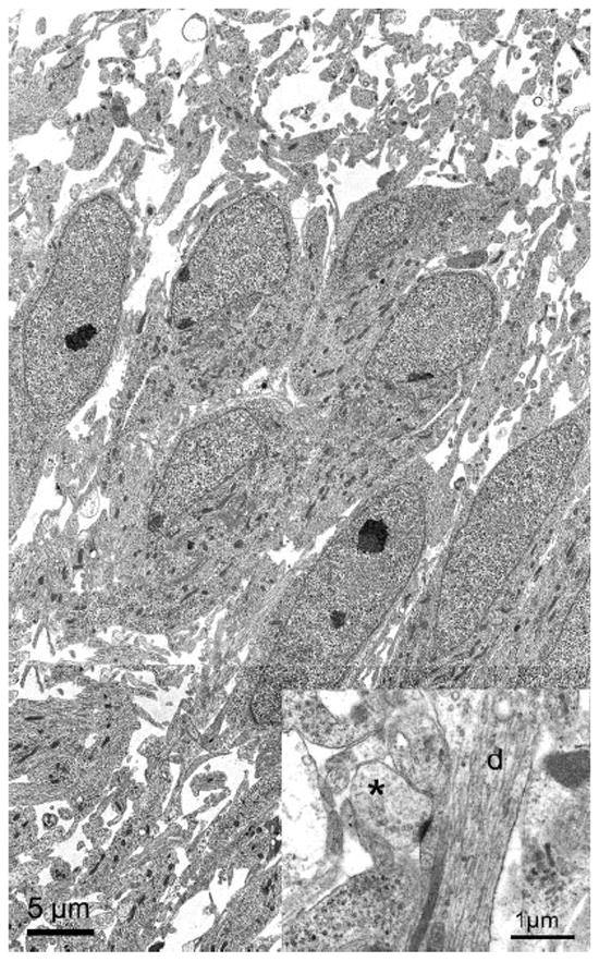
Electron micrographs obtained from NL at E18. Low-power electron micrographs of NL showed the neurons to be tightly packed in the center of the nucleus. In this region of NL, neurons exhibited polarized dendrites that project primarily in the dorsoventral dimension. Dendrites can be readily identified at larger magnification (inset, d). A synapse (asterisk) can be clearly identified making contact with an NL dendrite. The synapse is from an adjacent section.