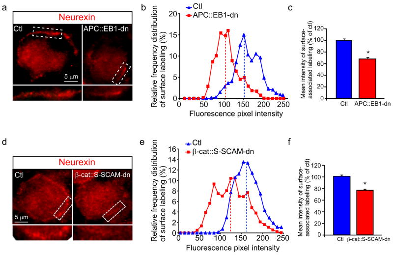
Figure 7. Neurexin surface clusters are decreased on presynaptic terminals that contact postsynaptic neurons expressing APC::EB1-dn or β-cat::S-SCAM-dn.
Micrographs (a,d) of immunofluorescence double-labeled E13 CG frozen sections showing that Nrx surface clusters (red, a,d) are decreased on presynaptic terminals that contact APC::EB1-dn (a) and β-cat::S-SCAM-dn (d) neurons as compared to Ctl neurons. Insets, two-fold magnification views of boxed regions. Nrx staining shows shifts to lower pixel intensity levels (b,e) as well as 31.5% and 22.6% reductions in mean intensity levels (c,f) at synapses on APC::EB1-dn (b,c) and β-cat::S-SCAM-dn (e,f) expressing neurons, respectively, relative to Ctl neurons. (APC::EB1-dn: *p<6.9 × 10⁻⁹, Student t-test, n=21 DN and 14 Ctl neurons; β-cat::S-SCAM-dn: *p<5.2×10⁻²² Student t-test, n=22 DN and 21 Ctl neurons). Dashed vertical lines indicate the median intensity values (b,e). Bars represent the mean ± SEM (c,f).

