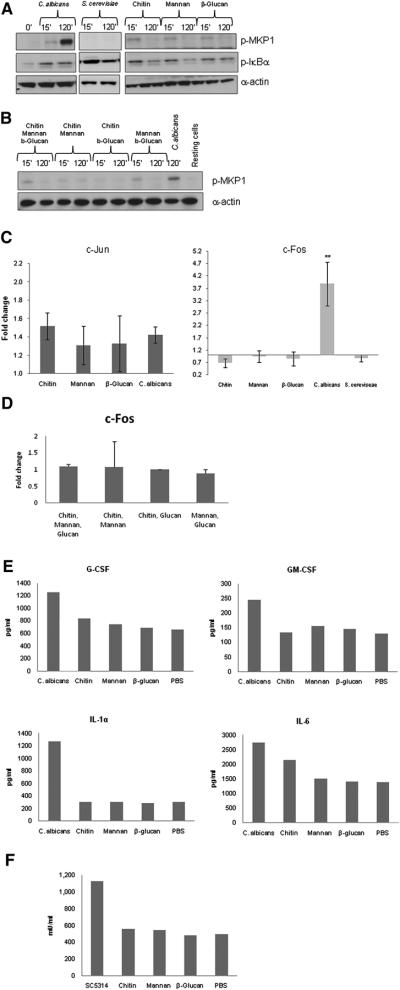
Figure 5. Activation of MKP1 and c-Fos by C. albicans and S. cerevisiae and epithelial responses to fungal cell wall components.
(a–b) Phosphorylation of MKP1 and IκBα after 2 h or (c–d) activation of c-Jun and c-Fos DNA binding activity after 30 min and 3 h of infection, respectively, with either C. albicans or S. cerevisiae or treatment with 2 μg/ml C. albicans chitin, 50 μg/ml C. albicansN- and O- mannan or 100 μg/ml β-glucan microspheres (individually or in combination) for the same time periods. Only C. albicans activates MKP1 and c-Fos. (e) Moiety-induced cytokine production after 24 h as measured by multiplex microbead assay. (f) Moiety-induced damage as measured by LDH release after 24 h. Doses for (b–d) were the same as those for (a). All experiments are (a–b) representative of or (c–e) the mean of at least three independent experiments. An MOI of 10 was used for both species. * = p<0.05, ** = p<0.01.

