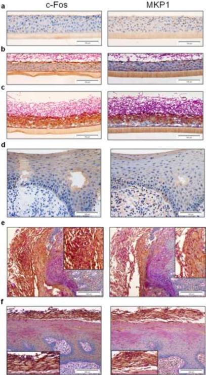
Figure 7. Expression of c-Fos and MKP1 in oral epithelium.
(a) Resting expression of c-Fos and MKP1 in oral RHE. Upregulation of c-Fos and MKP1 expression is associated with contact with Candida hyphae at (b) the surface after 4 h and (c) throughout the epithelial layer when hyphae penetrate and invade at 24 h (dark brown staining). (d) Resting expression levels of c-Fos and MKP1 in control biopsies of human oral epithelium. (e and f) Increased expression of both c-Fos and MKP1 in two different oral biopsies with Candida infection (panel e: left and panel f: top; dark brown staining). Insets in (e) and (f) show an enlarged view of the region of Candida infection in each respective biopsy.

