Figure 5.
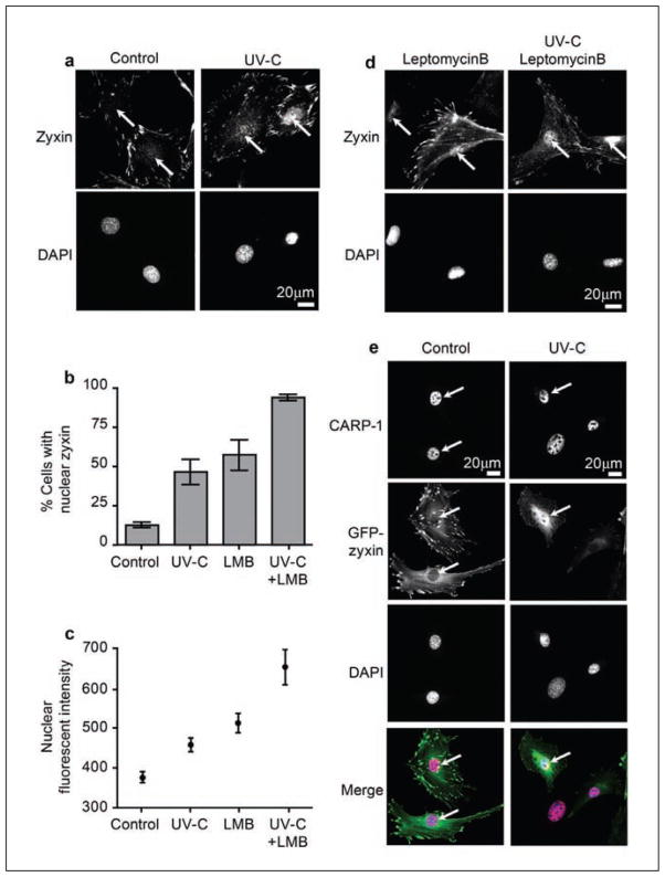
Zyxin transits to the nucleus in response to UV irradiation. (a) Indirect immunofluorescence microscopy of zyxin localization in untreated or UV-C–exposed cells. (b) Quantitative analysis of the percentage of cells with detectable nuclear zyxin in each treament condition. (c) Quantitative analysis of nuclear-localized zyxin by indirect immunofluorescence. (d) Indirect immunofluorescence microscopy of zyxin localization in untreated or UV-C exposed cells in the absence or presence of Leptomycin B (20 nM, 6 hours). (e) CARP-1 is concentrated in cell nuclei both before and after UV exposure. In cells programmed to express GFP-zyxin, which localizes normally to focal adhesions, zyxin is observed to accumulate in the nucleus and co-localize with CARP-1 upon UV-C exposure. For Merge images, zyxin (green), CARP-1 (red), and DAPI (blue) merges to white when all 3 signals overlap. Arrows indicate the position of the nucleus.

