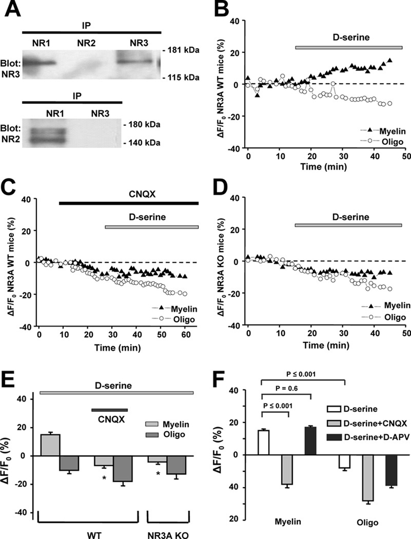
Figure 1.
Presence and physiological response of NR1/NR3 receptors in optic nerve myelin. A, Co-immunoprecipitation from the myelin fraction using antibodies against NMDAR subunits. Antibodies used for the precipitation are indicated above the lanes. The membrane was probed with an anti-NR3 antibody (upper blot) and a pan-NR2 antibody (bottom blot), yielding evidence for NR1/NR3 but not NR2/NR3-containing complexes. Molecular weight markers are indicated at right. B, Addition of 500 µM d-serine induced a rise in Ca2+ in myelin from WT mice within minutes of application, an effect not observed in oligodendrocyte cell bodies. C, Rise in myelinic Ca2+ in response to d-serine was completely abrogated by 40 µM CNQX. D, In NR3A KO mice, d-serine failed to induce a rise in myelinic Ca2+. E, Bar graph summarizing mean intensity change in fluorescence of the Ca2+-sensitive dye Xrhod-1 in myelin and oligodendrocyte somal cytoplasm of WT and NR3A KO mice after application of d-serine (**p ≤ 0.001 vs. WT myelin exposed to d-serine). F, In rat, as in mouse, d-serine evoked significant increases in myelinic but not oligodendrocyte cell body Ca2+. The myelinic Ca2+ response was completely blocked by CNQX but unaffected by d-APV (200 µM).

