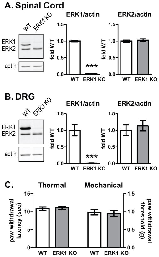
Figure 1. Disruption of mapk3 eliminates ERK1 expression in spinal cord and dorsal root ganglion without affecting heat or light touch thresholds.
A. Spinal cords were isolated from WT (n=13) and ERK1 KO (n=14) littermates and analyzed by Western blot for ERK1/2 and the loading control, actin. To quantify the intensity of ERK bands, integrated intensities of each isoform were divided by actin integrated intensities, and plotted as fold WT. B. DRG from WT (n=7) and ERK1 KO (n=8) were dissected and analyzed by quantitative Western blot as in A. In both spinal cord and DRG, there is a dramatic reduction in ERK1 expression without a significant change in ERK2 protein levels. C. No differences were observed in heat thresholds obtained by applying radiant heat to the hindpaws of WT (n=10) and ERK1 KO (n=10) littermates using a Hargreaves-style apparatus. No differences were observed in hindpaw mechanical withdrawal thresholds obtained with von Frey filaments from WT (n=14) and ERK1 KO (n=13) littermates. Error bars indicate S.E.M. (unpaired t-test; *** p<0.001).

