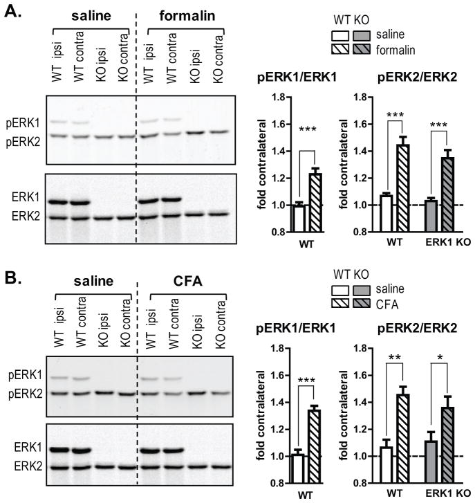
Figure 7. Inflammation-induced ERK2 phosphorylation is not elevated in ERK1 KO mice.
A. ERK1 KO and WT littermates were injected subcutaneously into the plantar surface of the hindpaw with saline or 3.5% formalin (n=6 per group) and sacrificed 3 minutes later. The dorso-ventral extent of L3–L6 spinal cord was isolated and processed for pERK1/2 and ERK1/2 quantitative Western blot analysis. Ipsilateral pERK/ERK intensities were normalized to contralateral intensities from the same subject, and data were graphed as fold contralateral. Formalin injection led to significantly elevated pERK1 and pERK2 in WT mice. In ERK1 KO mice, the formalin-induced increase in pERK2 was similar to WT. B. ERK1 KO and WT littermates were injected subcutaneously into the plantar surface of the hindpaw with saline or CFA (n=5–6 per group). After 30 minutes, the dorsal halves of L3–L6 spinal cords were obtained and analyzed for pERK1/2 and ERK1/2 by Western blot as above. As with formalin injection, CFA injected WT mice had significantly elevated pERK1 and pERK2. Elimination of ERK1 had no significant impact on CFA-induced pERK2 elevation. Error bars represent S.E.M. (unpaired t-test or 2-way ANOVA with Bonferroni post-test;* p<0.05, ** p<0.01, *** p<0.001).

