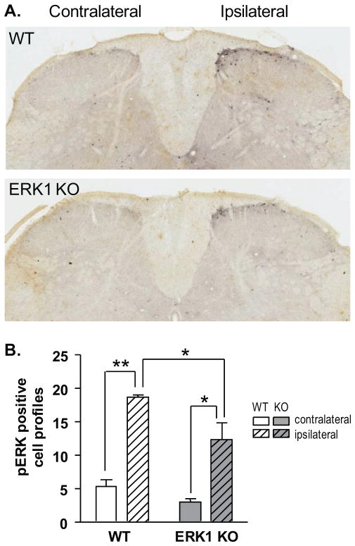
Figure 8. Noxious peripheral stimulation induces pERK1/2 immunoreactivity in superficial dorsal horn laminae despite the loss of ERK1.
A. Three minutes following subcutaneous intraplantar injection of 3.5% formalin, WT and ERK1 KO littermates were sacrificed and analyzed for pERK1/2 immunoreactivity in lumbar spinal cord. On the side of the spinal cord ipsilateral to paw injection, pERK1/2-positive cell profiles appear in the superficial lamina of formalin-injected WT and ERK1 KO mice. Diffuse staining is also apparent, possibly due to ERK1/2 phosphorylation in dendrites or afferent fibers. In both WT and ERK1 KO, there is no gross difference in the anatomic pattern of pERK1/2 staining. B. To quantify results from A., pERK1/2 positive cell profiles in laminae I–II were counted from 6 sections per animal with 3 animals per group. The mean number of cell profiles for each subject was then used to calculate the mean number for the entire group. Formalin stimulation significantly increases pERK1/2-positive cell profiles in both WT and ERK1 KO littermates (2-way ANOVA with Bonferroni post-test; * p <0.05, ** p<0.01). Error bars reflect S.E.M.

