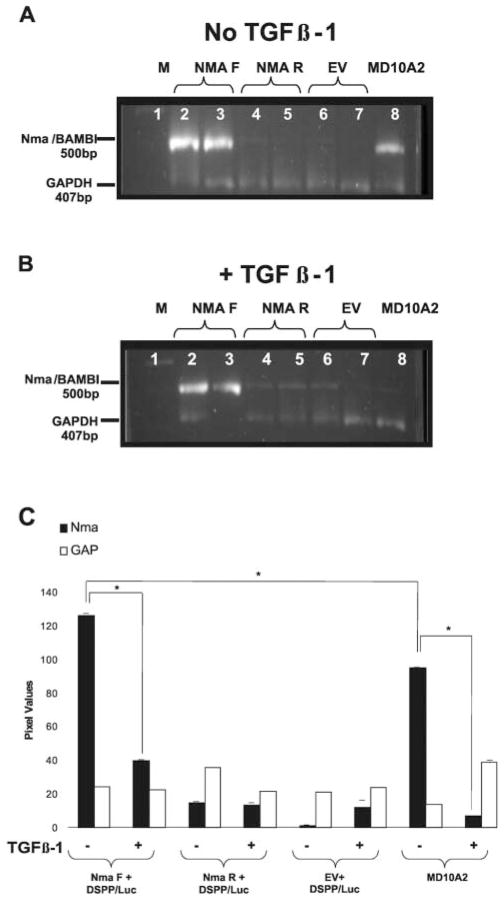
Figure 1.
RT-PCR amplification of Nma/BAMBI expression (500 bp) vs. GAPDH (407 bp) transcripts from MD10-A2 cells transiently co-transfected with an Nma/BAMBI-V5-His expression construct and a DSPP-Luc reporter construct. Amplification of transfected MD10-A2 mRNA from cells grown in control media (A) or supplemented with TGFβ-1 (B). Lane 1: 100-bp marker. Lanes 2 & 3: transfection with NMA/BAMBI-V5-His in the forward orientation (Nma F) and DSPP promoter-luciferase construct (DSPP-Luc). Lanes 4 & 5: transfection with NMA/BAMBI-V5-His in the reverse orientation (Nma R) and DSPP-Luc. Lanes 6 & 7: transfection with the empty vector (EV) and DSPP-Luc. Lane 8: non-transfected MD10-A2 cells. Increased Nma/BAMBI expression (lanes 2 & 3) compared with non-transfected MD10-A2 cells (lanes 8) is evident regardless of TGF® supplementation (A, B). (C) Semi-quantitative analysis of RT-PCR Nma/BAMBI expression data presented in panels A & B. Cells treated with TGF®-1 show a statistically significant reduction in Nma/BAMBI expression compared with untreated cells within the Nma F+DSPP-Luc transfected group and within the MD10-A2 control group (n = 3; p = 0.05; ± SD). Nma F+DSPP/Luc transfected cells had statistically significantly increased Nma/BAMBI expression compared with MD10-A2 control cells (n = 3; p = 0.05; ± SD).

