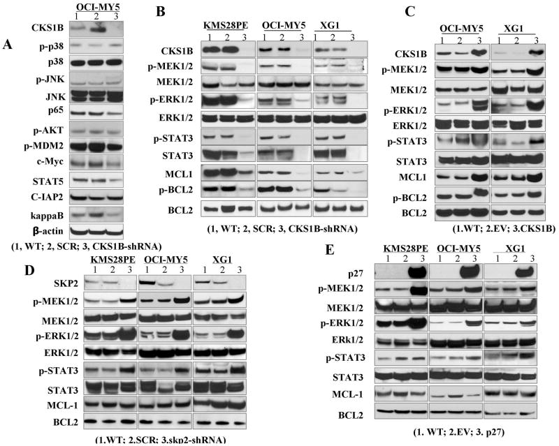
Fig. 3. CKS1B-over-expression activates STAT3 and MEK/ERK signaling pathways.
CKS1B was knocked down in KMS28PE, OCI-MY5 and XG-1 cells. Cells were cultured for 72 hours and cell lysates were prepared. (A) CKS1B-induced signaling pathways were screened by Western blots in CKS1B-knockdown (CKS1B-sh) OCI-MY5 cells using the indicated antibodies. (B) Protein levels of p-STAT3, p-MEK1/2, p-ERK1/2 and p-BCL2 were examined in KMS28PE, OCI-MY5 and XG1 cells after CKS1B-knockdown. Wild-type (WT) and Scramble (SCR)-transfected cells were used as controls and β-actin was used as loading control. (C–E) Protein levels of p-STAT3, p-MEK1/2, p-ERK1/2 and p-BCL2 were examined by Western blot analysis in CKS1B-transfected, SKP2-silenced, and p27 Kip1-transfected MM cell lines of OCI-MY5 and XG-1 cells, respectively. WT and EV cells were used as controls and β-actin was used as loading control.

