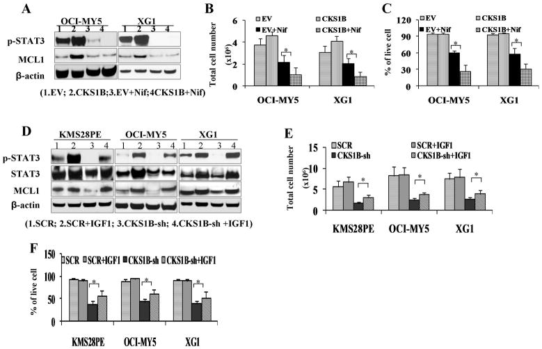
Fig. 4. Activation of STAT3 is involved in CKS1B-mediated myeloma cell growth and survival.
(A) 1×10⁶ CKS1B- transfected OCI-MY5 and XG-1 were treated with 10nM Nifuroxazide (Nif) for 48 hours, and cell lysates were prepared. Protein levels of p-STAT3 and MCL1 were analyzed and showed decreased in Nif treated MM cells by Western blots. (B) Cell growth and (C) cell survival were evaluated. MM cells treated with Nif showed clearly more cell growth inhibition (OCI-MY5: 2.18M vs. 1.04M, and XG-1: 2.07M vs. 0.90M) and more cell death (OCI-MY5: 60% vs. 27%, and XG1: 58% vs. 31) in CKS1B over-expressed cells. Untreated and EV cells with or without Nif treatment were used as controls. (D) Western blots showed IGF-1 increased p-STAT3 and MCL1 in CKS1B silenced MM cells. 1×10⁶ OCI-MY5 and XG-1 after CKS1B-knockdown (CKS1B-sh) were treated with 100 ng/mL IGF-1 for 72 hours and p-STAT3 and MCL1 protein levels were analyzed. Untreated CKS1B-sh cells and SCR cells with or without IGF-1 treatment were used as controls. (E) Cell growth and (F) cell death were evaluated. IGF-1 can partially rescue CKS1B knockout induced cell growth inhibition (KMS28PE: 1.74M vs. 2.88M, OCI-MY5: 2.40M vs. 3.64M, and XG1: 2.60M vs. 3.86M) and apoptosis (KMS28PE: 36% vs. 56%, OCI-MY5: 43% vs. 61%, and XG1: 39% vs. 53%). Untreated cells and SCR cells with or without IGF-1 treatment were used as controls. Results were expressed as Mean ± SD of three independent experiments (*p <0 .05).

