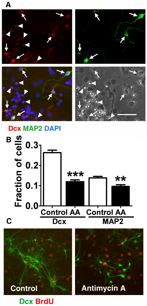
Fig. 4.
Immature Dcx+ neurons are selectively vulnerable to mitochondrial inhibition. A. Cultures treated with antimycin A, 2 μM for 16 h, stained with an immature neuron marker Dcx (red) and a marker for more mature neurons MAP2 (green). Cell nuclei are counterstained with DAPI (blue). The cultures were stained after 16 h of antimycin A treatment, compared to the 24 h time point used for quantification to capture the residual Dcx staining that is essentially gone after 24 h. Cells expressing both Dcx and MAP2 cell markers (arrows) showed better preserved morphology and Dcx staining compared to Dcx-only expressing cells (arrowheads). Scale bar is 50 μm. B. Fractions of Dcx+ and MAP2+ cell (relative to the total cell number) in control and antimycin A (2 μM, 24 h) treated cultures. The data are representative of three independent experiments with at least 700 cells per condition in each experiment .mean±SEM (**p<0.01 compared to control MAP2, ***p<0.001 compared to control Dcx). C. Only a small fraction of Dcx+ cells (green) were proliferating after 8-9 days of differentiation, as demonstrated by co-labeling with BrdU (red). The fraction of proliferating BrdU cells was not significantly changed by 24 h antimycin A treatment.

