
Figure 10. A scheme that summarizes the subcellular organization of Dmoesin and the major signaling proteins in the rhabdomeric membrane.
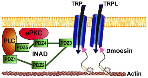
TRP anchors the INAD signaling complex, which includes PLC and eyePKC (ePKC), to the plasma membrane via the PDZ3 domain of INAD. The NH2-terminal region of Dmoesin molecules (arrowheads) bind either directly or through a PDZ adaptor protein to the TRP and TRPL channels, whereas the COOH-terminal region is bound to the actin cytoskeleton. The phosphorylated site of T559 is indicated by an asterisk.