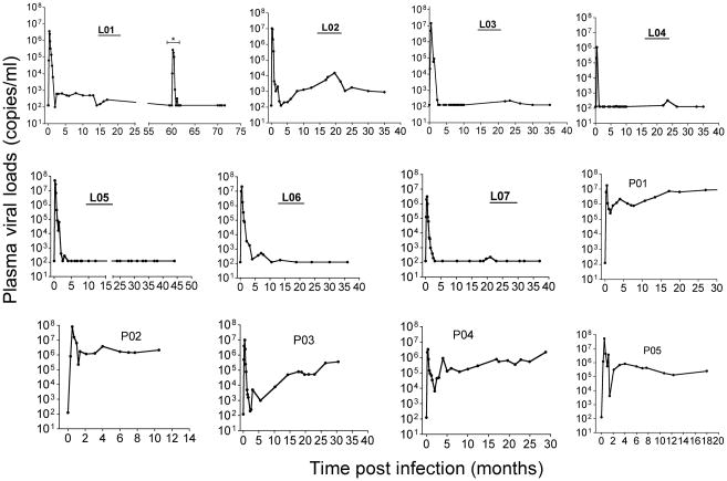
Figure 1.
Plasma viral loads in SIVmac-infected rhesus macaques of Chinese-origin. Animal numbers of LTNP animals are shown in bold and underlined. Animal L01 had a transient peak of viremia ~ 60 months after infection due to experimental CD8+ T cell depletion (shown by a * in the figure), but viremia was undetectable thereafter. The limit of detection was 125 copies/ml by bDNA assay. Intestinal biopsies were taken after viral set point at the chronic phase of SIV infection ranging from 10 months (P02) to 6 years (L01).

