
Figure 5.
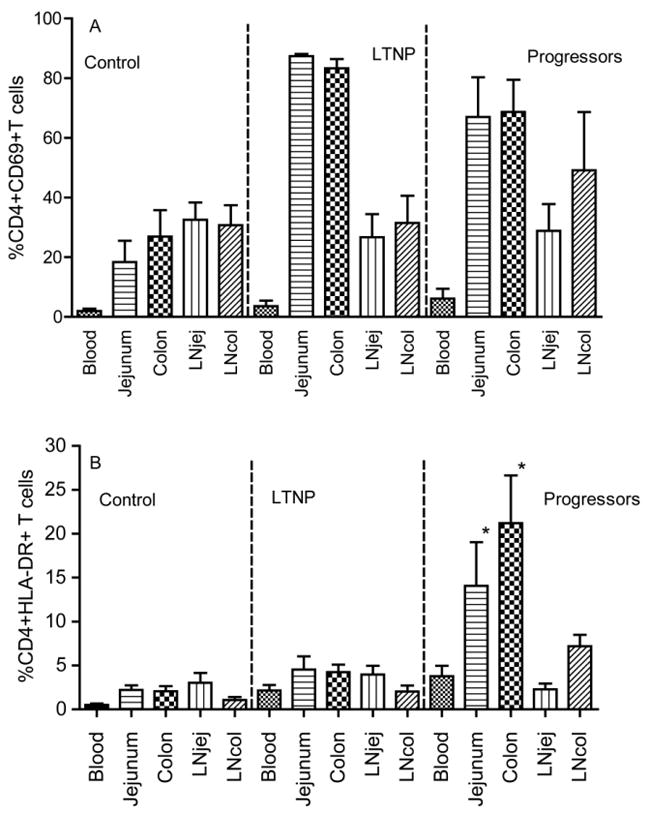
Comparison of T cell activation in different tissue compartments in normal Chinese rhesus macaques, LTNP and progressors as determined by early activation marker CD69+ on CD4+ T cells (A) and HLA-DR expression on CD4+ T cells (B). *: P < 0.05 compared with peripheral blood within the same group.