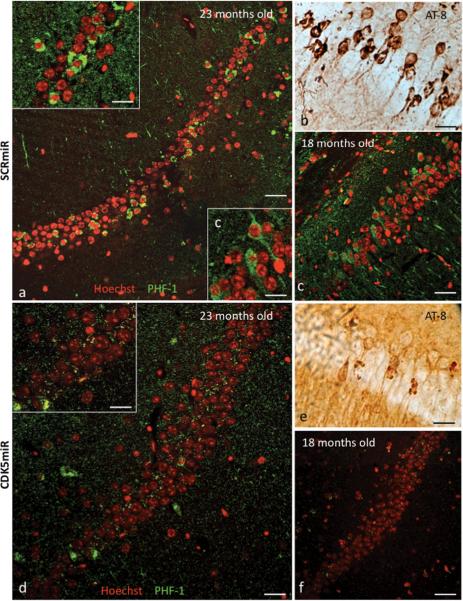
Figure 5. CDK5miR decreases phosphorylated tau and neurofibrillary tangles in 3xTg-AD mice.
a) Phospho-tau (PHF-1) immunofluorescence showing neurofibrillary tangles (NFTs) positive cells and b) phospho-tau (AT-8) immuoreactive cells present in the CA1 area of hippocampus of 23 month old 3xTg-AD mice three weeks post-injection with AAV-SCRmiR. c) PHF-1 immuofluorescence showing positive cells in CA1 in hippocampus of 18 month old 3xTg-AD mice, three weeks after hippocampal injection with AAV-SCRmR. d) PHF-1 immunofluorescence and e) AT-8 immunoreactive cells in the CA1 area of 23 months old and f) 18 months old 3xTg-AD mice three weeks after injection with CDK5miR. n=6, Red pseudo-color: nucleus staining with Hoechst, green pseudo-color: PHF-1 IF, Alexa 594. 60×, Bar: 50 μm; Inserts a, c, d: 100×, scale bar: 20 μm.

