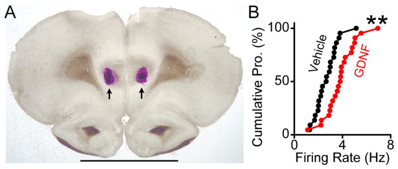
Figure 5. Intra-NAc infusion of GDNF leads to an increase in spontaneous firing rate of PFC-projecting VTA neurons.

A–B, GDNF (10 μg/ 2μl) was bilaterally infused into the NAc 7-11 days following intra- PFC infusions of DiI. VTA slices were prepared 12 hrs after GDNF infusion, and the spontaneous firing of DiI-labled PFC-projecting neurons was measured. A, A representative coronal section confirming the injection sites within the PFC. Arrowheads indicate DiI deposit. Scale bar, 0.5 mm B, Cumulative probability plot comparing spontaneous firing rates of individual neurons in slices from vehicle- (black circles) and GDNF- (red circles) treated rats. ** p < 0.01 vs. vehicle by Kolmogorov-Smirnov test, n = 22 cells from 5 rats for each group.
