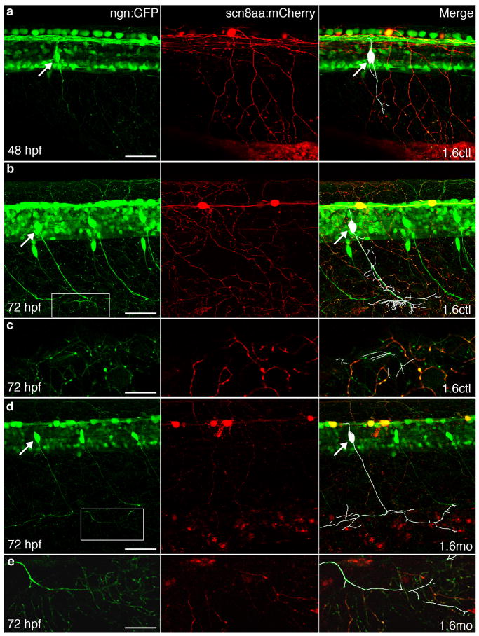
Figure 4.
RB and DRG peripheral arbors overlap by 72 hpf in 1.6 control and morphant embryos. In all panels, GFP (green) in Tg(−3.4neurog1:GFP) embryos labels both RB and DRG axons and cell bodies, whereas stochastic labeling with mCherry allows visualization of peripheral arbors (red) of individual RB cells. In the left merged panels, white traces the processes of the DRG neuron. White arrows indicate cell bodies of DRG neurons. A, In 48 hpf embryos injected with 1.6ctl, DRG axons that run next to the notochord have not yet reached the periphery and do not overlap with cutaneous RB peripheral processes. B,C,D,E, By 72 hpf, in embryos injected with either 1.6ctl (B,C) or 1.6mo (D,E), DRG neurons extend processes into the skin where they overlap with RB cutaneous processes. Panels C and E show 8 μm confocal slices of the regions boxed in B and D, respectively. Scale bars, 50 μm: A, B and D; 20 μm: C, E.

