
Fig. 1.
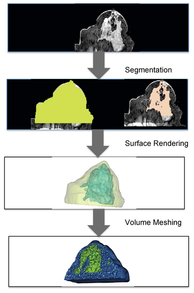
A schematic showing steps from medical image data to obtaining a volumetric mesh for computation with examples from breast data. These steps have to be routinely performed before image reconstruction can be done for 3D multi-modality optical imaging. Methods for image segmentation vary between applications; here thresholding and region-growing techniques were applied for breast tissue. Surface rendering is automatically generated by many open source softwares, but getting a reliable volume mesh can be time-consuming and more difficult to automate.