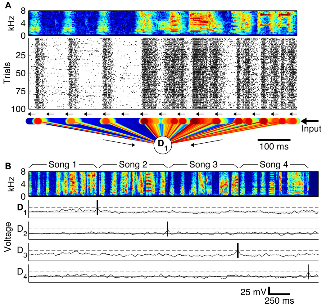
FIG. 1.
The spike pattern recognition model uses time-varying properties of the neural responses to recognize spiking patterns, and consists of a linear chain of neurons. Sensory input (here from field L) feeds into one end of the chain, propagating along the chain using synaptic delays, turning the temporal spiking pattern into a spatial activation of chain neurons. A: Bird song (spectrogram) elicits auditory responses (spike rasters), leading to excitatory (red) and inhibitory (blue) synapses from 298 sequentially-connected chained neurons (small circles) that each connect to a detector (D1). The spectrogram shows the power (color, red: high power, blue: low power) in different frequency bands (y-axis) as a function of time (x-axis), while the rasters show one field L neuron’s responses to 100 repeated presentations (y-axis) of the above stimulus, with each tick mark representing an elicited action potential. B: When the chain recognition circuit (with four detectors D1–D4 shown) is fed input from the field L neuron in response to the concatenation of four different songs (spectrograms), each detector fires only at the end of the correct trained song (D5–D20 not shown).

