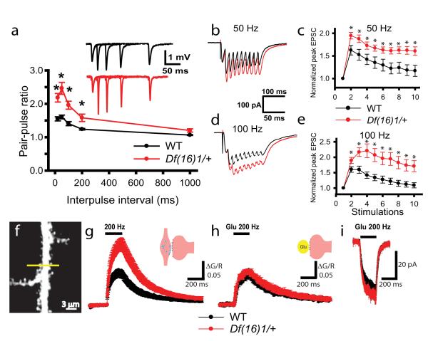
Figure 3. Presynaptic function is affected by the Df(16)1 microdeletion in mature mice.
(a) PPF is enhanced in mature Df(16)1/+ mice. Mean fEPSPs as a function of the interstimulus interval (ISI) between the first and second fEPSPs evoked at CA3-CA1 synapses of WT and Df(16)1/+ mice. Inset shows representative traces of pairs of fEPSPs evoked with 20-, 50-, 100-, or 200-ms ISIs. (b-e) Short-term plasticity is enhanced in mature Df(16)1/+ mice. Typical EPSC traces (b, d) and mean peak amplitudes of EPSCs (c, e) evoked by 50-Hz (b, c) or 100-Hz (d, e) synaptic stimulations in slices from mature WT and Df(16)1/+ mice (* p<0.05). (f-h). Calcium transients evoked by synaptic stimulation but not by TGU are stronger in Df(16)1/+ mice. (f) A TPLSM image of a CA1 pyramidal neuron dendrite. The line represents the direction of the line scan. (g, h) Mean normalized Fluo 5F fluorescence (ΔG/R) in dendritic spines of CA1 neurons as a function of time with 40 synaptic (g) or TGU (h) stimulations delivered at 200 Hz. (i) Mean EPSCs evoked by 40 pulses of TGU at 200Hz at dendritic spines.

