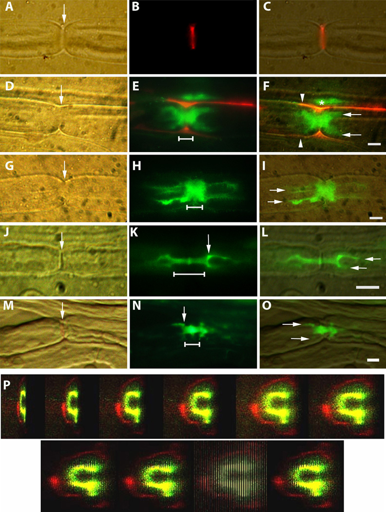
Fig. 1.
A–O., Hoffman, fluorescence and merged images showing penetration of dextran tracers into node/paranode regions of sciatic nerve myelinated fibers. Scale bars = 5microns. The node of Ranvier is indicated by arrows in A, D, G, J and M. The linear extent of ‘paranodal bars’ is indicated by horizontal lines in E, H, K and N.
A–C. Live injection. 70K tracer (red) fills the nodal slit (arrow).
D–F. Live injection. 70K tracer (red) outlines a nerve fiber (E and F). Between the red outlines (arrowheads, F), 3K tracer (green) has penetrated from the node into the fiber symmetrically forming a thick paranodal ‘bar’ just beginning to give rise to the tines (arrows, F) of ‘hairpins’ at both ends. Some 3K tracer above this fiber (*) has been trapped between it and a 2nd fiber whose outline is visible above.
G–I. Live injection. 3K tracer (green) forms a paranodal ‘bar’, which widens to form conspicuous ‘hairpins’ (arrows) symmetrically on both sides of the node.
J–L. Fixed nerve exposed to 3K tracer (green), which forms a narrow paranodal ‘bar’ (line, K) that is slightly wider just at the node. At both ends, the bar widens in a ‘shoulder’ region (arrow, K), where it forms ‘hairpins’ that extend into the intermodal periaxonal space.
M–O. Fixed nerve exposed to 70K tracer (green), which forms a short ‘bar’ (line, N) that gives rise to square ‘shoulder’ regions (arrow, N) at each end and the beginnings of ‘hairpin’ tines (arrows, O).
P. Live injection. Confocal Z stack rotated to show dark core (internodal axon), surrounded by 3K dextran (green) in the periaxonal space. That in turn is surrounded by a dark ring (compact myelin) and outside of that a thin red ring (70K dextran) along the outside of the fiber. The 9th image in the series shows the confocal slices end on.

