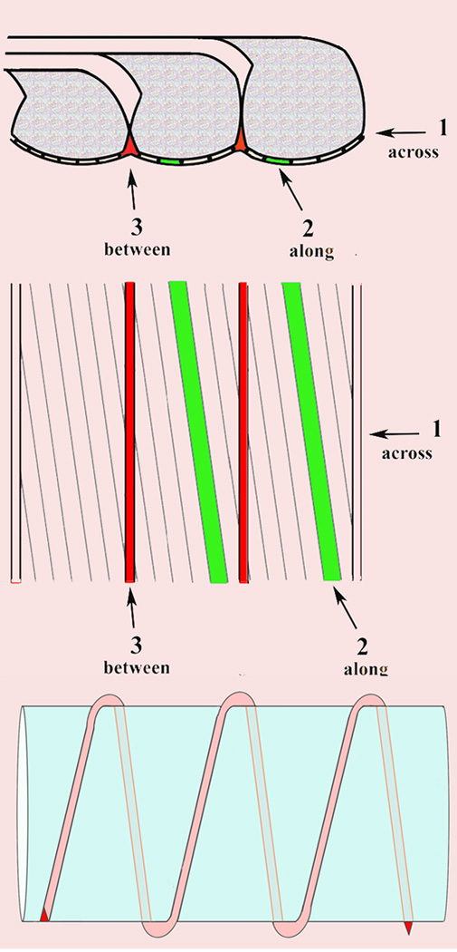
Fig. 3.
Schematic representations of possible pathways of diffusion
Top. Side view of paranodal loops.
Middle. Tangential view. Pathway 1 goes through the junctional cleft across the transverse bands. Pathway 2 also goes through the junctional cleft but passes along the transverse bands at ~8 degrees from transverse. Pathway 3 does not go through the junctional cleft but passes between paranodal loops.
Bottom. Longitudinal view of pathway 3 winding helically around an axon.

