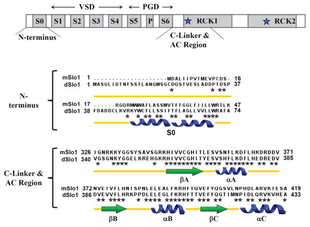
Figure 1. Schematic of BK channel structure.
Top: S0–S6 are transmembrane segments, RCK1 and RCK2 are located in cytoplasm. VSD: voltage sensing domain; PGD: pore-gate domain. * identifies conserved residues and ★ represents the location of Ca2+ binding sites. The sequence and secondary structure of the N-terminus including S0 and the C-Linker and AC region are shown below.

