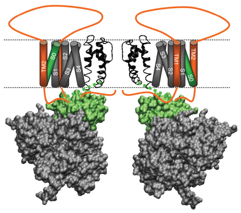
Figure 9. A structural model for the β2 subunit modulation.

Two opposing mSlo1 and β2 (orange) subunits of BK channels are shown where cartoons for the VSD and S0 segment are constructed around the pore of the MthK channel (PDB ID: 1LNQ) (Jiang et al., 2002). The gating ring of the BK channel (PDB ID: 3MT5) (Yuan et al., 2010) is aligned to the MthK channel using UCSF Chimera v1.4.1. VMD v1.8.7 was used to show the aligned structure in cartoon and surface representation. The helices in the VSD are shown as cylinders and are positioned according to the KV1.2 channel. The transmembrane (TM) segments of the β2 subunit are positioned according to Zakharov et al (Zakharov et al., 2009). Green colored regions in the Slo1 subunit (S0, C-Linker and AC) are involved in the β2 modulation of Ca2+ sensitivity.
