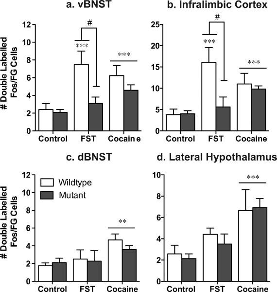
Figure 5. CREB mutant mice exhibit a decrease in stress-activated cells that project to the VTA.
While wildtype mice exhibit an increase in Fos activated cells that project to the ventral tegmental area [doubled labeled Fos/Flourogold (FG) cells] in the ventral BNST (a) and the Infralimbic cortex (b), CREBαΔ mutant mice do not. Although no differences were seen between the genotypes in response to acute cocaine, there was an increase in double-labeled cells in two regions that did not show stress-induced alterations, the dorsal BNST (c) and the lateral hypothalamus (d). *p<.05, **p<.01, ***p<.001 as compared to control; #p<.05 pair-wise comparison wildtype FST vs. mutant FST.

