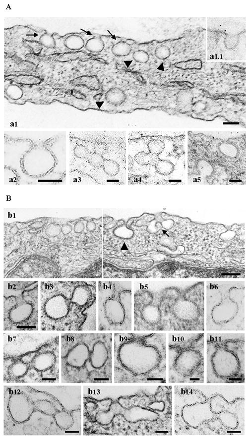
Figure 2. Expression of the SH3A of ITSN-1 in cultured ECs causes abnormal caveolae morphology.

(A) The electron micrographs show a segment of a control ECs with omega-shaped caveolae open to the extracellular environment through short necks (a1 arrows, a2) or directly (inset a1.1), and caveolae as apparently detached spherical structures in the cytosol (Fig. 2A, a1, arrowheads). Clusters of interconnected caveolae are shown in a3, a4. Two clathrin-coated pits open on opposite sides of the PM are shown in a5. Bars: 50 nm. (B) Electron micrographs showing highly magnified caveolar profiles with unusual morphology. Caveolae (b1, b4–b6) and clathrin-coated pits and vesicles (b1, arrowhead, b2) with elongated necks were frequently observed. The micrographs in panels (b7 – b11) show staining-dense collars encircling the caveolae necks. Myc-SH3A expression causes unusual caveolae clustering (b1, arrow, b12 – b14). Bars: 100 nm (b1); 70 nm (b2 – b6); 50 nm (b7, b8, b12 – 14); 25 nm (b9–b11).
