
Figure 4. ICa,Cl as marker for Ca2+ cyt levels.
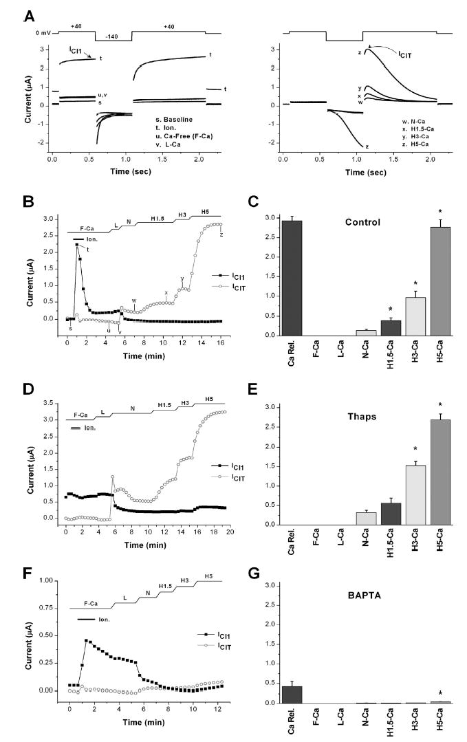
ICa,Cl was recorded from control cells (untreated), cells treated with thapsigargin (2 μM, 3 h), or injected with 500 μM BAPTA to estimate store Ca2+ load and the levels of Ca2+ influx. ICa,Cl provide endogenous reporters of Ca2+ release from stores (ICl1) and Ca2+ influx from the extracellular space (IClT) as described in the text. (A) Voltage protocol and representative current traces of the Ica,Cl. ICl1 is a sustained current recorded upon depolarization to +40 mV (trace t), whereas IClT is a transient current detected only when the +40 mV pulse is preceded by a hyperpolarization step to −140 mV (traces x–z). Note that at high 5 mM Ca2+ levels, Ca2+ influx at −140 mV activates an inward Cl− current (trace z). The current traces shown are from the control oocyte in B. The time at which each trace was obtained is indicated in B. (B–G) Oocytes were incubated in Ca2+-free Ringer (F-Ca) and treated with ionomycin to release store Ca2+. The levels of ICl1 induced in response to ionomycin provide a measure of store Ca2+ load. ICl1 (squares) is plotted as the maximal current at the end of the +40 mV pulse as indicated by the arrow in A (left). After store depletion oocytes were sequentially exposed to solutions containing the indicated Ca2+ concentration: L (50 μM Ca2+), N (0.6 mM Ca2+), H1.5 (1.5 mM Ca2+), H3 (3 mM Ca2+), and H5 (5 mM Ca2+). Store depletion activates Ca2+ influx through the SOCE pathway, which activates IClT. IClT (circles) is plotted as the maximal current during the second +40 mV pulse as indicated by the arrow in A (right). B, D, and F show the time course of ICl1 and IClT in control, thapsigargin, and BAPTA-treated cells, respectively. The time of solution changes and ionomycin (Ion.) addition are indicated above each panel. C, E, and G show statistical analysis of ICl1 and IClT. ICl1 levels were significantly different in the three treatments (P ≤ 0.0041, n = 5–7). No ICl1 was detected in the thapsigargin treatment indicating complete Ca2+ store depletion. For IClT in each panel the asterisks indicate significantly different means: (C) Con, P ≤ 0.015, n = 5; (E) Thaps, P ≤ 0.00012, n = 7; (G) BAPTA, P = 0.00022, n = 6.