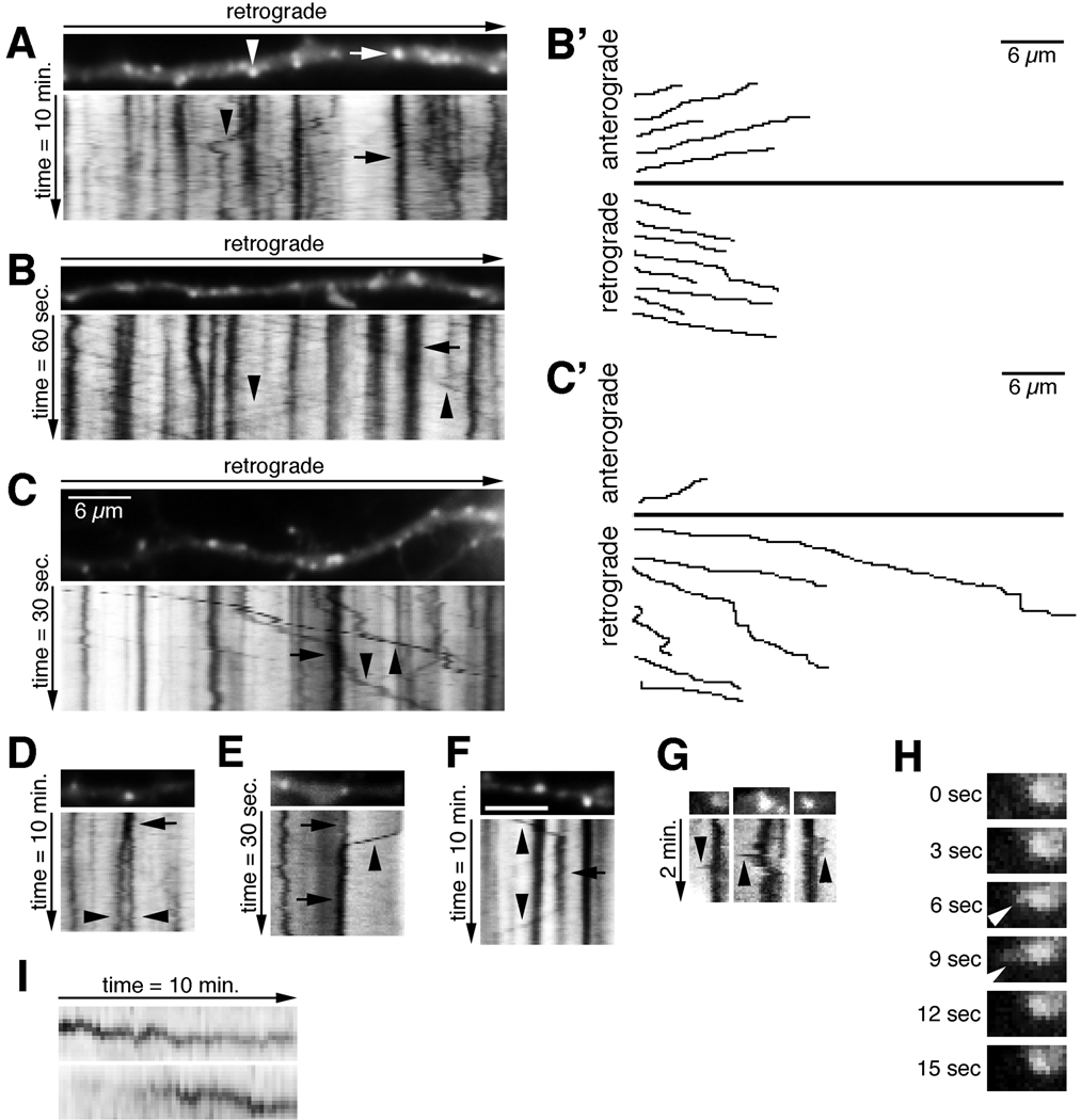
Figure 2. EHD1-positive endosomal compartments in neurons are dynamic.
(A–C) Neurons were transfected with cherry-EHD1. Images were captured with either 10 sec, 1 sec, or 0.5 sec frame rates and total duration of 10 min, 60 sec, and 30 sec, respectively. The first movie frame of a part of dendrite is shown with the corresponding kymographs underneath. The time dimension is shown on the y-axis, the retrograde direction is indicated above. Arrows indicate stationary vesicles and arrowheads motile vesicles. Scale bar is 6µm. (B’, C’) Trajectories of anterograde and retrograde moving vesicles from B and C. (D–F) Examples of fusion (E, F) and budding (D, F) of cherry-EHD1 vesicles during either 30 sec or 10 min time lapse. (G) Examples of dynamic shape changes of stationary vesicles in the soma, which include the temporary extensions of tubular domains (arrowheads; 3 sec frame rate). (H) Single frames of first time lapse from G magnified 2× are shown to illustrate example of tubular extension (arrowhead). (I) Examples of changes in fluorescence intensity of cherry-EHD1 vesicles during 10 minutes time-lapse.

