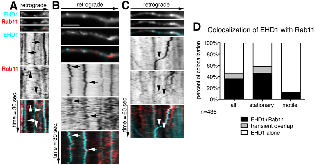
Figure 5. Dynamic co-localization of EHD1 with rab11-positive recycling endosomes.
(A–C) First frames of time-lapse of part of dendrite of neurons expressing cherry-EHD1 (cyan) and rab11-GFP (red) is shown with the corresponding kymographs underneath. The time dimension is shown on the y-axis, the retrograde direction is indicated above. Duration of time-lapse is 30 sec (A–B) or 60 sec (C), and frame rate 0.5 sec and 1 sec respectively. Scale is 6 µm. (A) Examples of stationary EHD1-positive vesicles (arrows) and motile rab11-positive vesicles (arrowheads). (B) Example of two stationary EHD1-positive vesicles that transiently colocalize with rab11. (C) The example of two motile rab11-positive vesicles (arrowheads), one of them colocalizes with EHD1. (D) Quantification of colocalization of EHD1 with rab11 in stationary and motile vesicles (n=436 endosomes). There are three classes of vesicles: EHD1-alone that are positive for only EHD1; EHD1+rab11 that are positive for both EHD1 and rab11, and “transient overlap” vesicles that contain EHD1 throughout time-lapse but rab11 only for part of the time-lapse.

