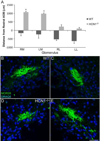
Figure 11.
In HCN1−/− mice MOR28 glomeruli change their location but have normal coalescence. A: The histogram shows the change in position relative to the first section containing AOB in the WT compared to HCN1−/− mice. B-E: OB sections from P0 mice labeled with MOR28 and DRAQ5. B-C: Left and right medial WT glomeruli, respectively. D-E: Left and right medial HCN1−/− glomeruli, respectively. RM, right medial; LM, left medial; RL, right lateral; LL, left lateral. Scale bar = 50 μm.

