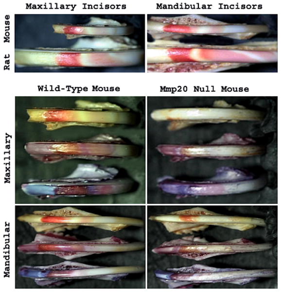
Figure 2. Staining of Incisors with pH Indicators.

A comparison of wild-type rat and mouse incisor banding pattern was made by use of methyl red staining (Top Panels). Note that for mandibular incisors, the rat has at least one more band of acidity than does the mouse. The four bottom panels show incisors from wild-type and Mmp20 null mice. For each of these panels the top incisor is stained with methyl red, the middle incisor is stained with bromophenol red and the bottom incisor is stained with resazurin. The staining pattern of the Mmp20 null incisors is distinctly different from the wild-type control and the areas of acidity are greatly reduced in the null mouse enamel.
