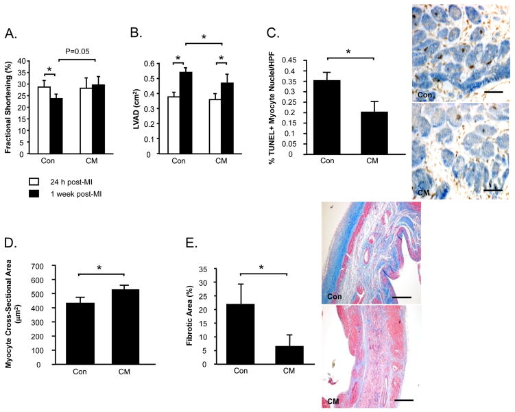
Figure 4. Intramyocardial injection of CM post-MI attenuates LV dysfunction and remodeling.
Echocardiography was performed to assess fractional shortening (A) and LV diastolic areas (LVAD) (B). Myocardial sections were stained to count TUNEL+ apoptotic myocytes (C). Masson’s Trichrome-stained sections were assessed for myocyte cross-sectional areas (D) and myocardial fibrosis (E). Data is presented as mean±sem. N=10-12/group. *P<0.05.

