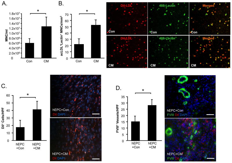
Figure 6. CM augments mobilization and homing of EPCs to the post-MI heart.
To examine the effect of CM treatment on EPC mobilization, peripheral blood was sampled from rats at the end of the 1 week study and total MNC were counted (A). acLDL+lectin+ EPCs cultured from the MNC fraction were counted (B). In a separate experiment, the effects of control medium or CM on DiI+ EPCs homing to the ischemic rat heart were examined. At 1 week post-MI myocardial sections were examined for the presence of DiI+ cells (C). Myocardial sections were stained to identify vWF+ vessels co localized with DiI+ EPCs (D). Data is presented at mean±sem. N=10–12/group. *P<0.05.

