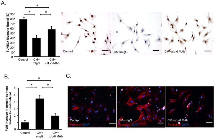
Figure 7. CM protects cardiac myocytes from apoptotic death and promotes myocyte hypertrophy.
The direct effects of control medium, CM+mouse IgG isotype control (mIgG) or CM+αIL-6 MAb on neonatal rat cardiac myocytes were examined. (A) Hypoxia-induced apoptosis was determined by TUNEL (brown). Blue: viable nuclei. Myocyte hypertrophy was determined by measuring total protein content, relative to DNA content (B) and phalloidin-staining of fixed cells (C). Data is represented as mean±SEM. Each condition was represented in triplicate. N=3 experiments. *P<0.05.

