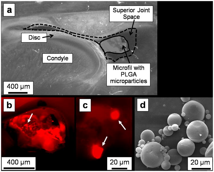
Figure 1.

Phase contrast and fluorescence microscopy images indicated successful injection of fluorescent-dye-loaded PLGA microparticles (MPs) into the superior joint space. In this representative unstained histological section from a rat euthanized immediately after injection (2.5× magnification), (a) the Microfil (solid outline) containing the PLGA MPs is visible within the superior joint space (dashed outline) via phase contrast imaging. A higher magnification (4×) fluorescent image of this same section confirms that (b) the fluorescent-dye-loaded MPs are contained within the Microfil (arrow points to an area with MPs), with (c) individual MPs (white arrows) visible within the Microfil at 40× magnification. A scanning electron micrograph (d) of the spherical PLGA microparticles is included for comparison.
