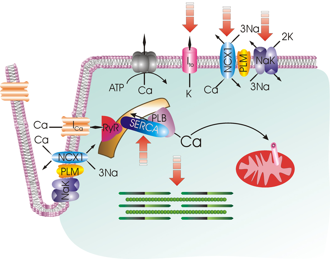
Figure 3. Cardiac excitation-contraction coupling.
Membrane depolarization is initiated by opening of the Na+ channel (not shown) with Na+ entry. Extracellular Ca2+ enters via L-type Ca2+ channel (ICa) and Na+/Ca2+ exchanger (NCX1), causing Ca2+ release from the ryanodine receptor (RyR) in the sarcoplasmic reticulum (SR). Ca2+ binds to troponin and initiates myofilament contraction. During diastole, Ca2+ is pumped back to the SR by SR Ca2+-ATPase (SERCA) under the control of phospholamban (PLB). A small amount of Ca2+ is also taken up by the mitochondrial Ca2+ uniporter. The amount of Ca2+ that has entered during systole is extruded by Na+/Ca2+ exchanger and to a lesser extent, by sarcolemmal Ca2+-ATPase. Na+ that has entered via Na+ channel and Na+/Ca2+ exchanger is pumped out by Na+-K+-ATPase. Repolarization is mediated by opening of K+ channels (only the transient outward K+ current Ito responsible for early repolarization is shown). Phospholemman (PLM) associates with and is an endogenous regulator of Na+/Ca2+ exchanger, Na+-K+-ATPase and possibly L-type Ca2+ channel. Na+/Ca2+ exchanger is depicted as operating in the forward mode (Ca2+ efflux) in the sarcolemma and reverse mode (Ca2+ influx) in the t-tubules. Broken arrows point to ion transporters, ion channels and myofilaments that are altered after myocardial infarction.

