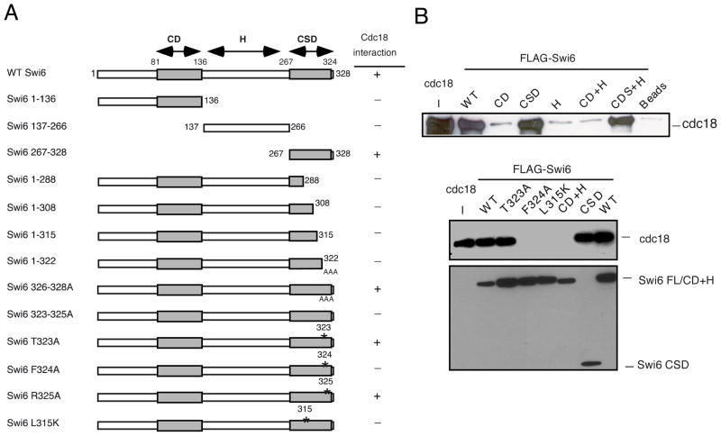
Fig 2. Mapping of the Swi6 interaction domain by in vitro binding.
(A) Schematic representation of the Swi6 protein and mutational analysis. The chromo domain (CD) is linked to the chromo shadow domain (CSD) through the hinge (H) region summarizes the inding results. (B) In vitro binding studies were performed by precipitation of bacterial recombinant His6FLAG-Swi6 wild-type or fragments or mutants with FLAG M2-agarose conjugate beads. Recombinant MBP-Myc Cdc18 wild-type was added to the beads and its binding capacity was measured by western blotting. Immunoblotting was performed with anti-Myc antibodies for Cdc18 or anti-FLAG antibodies for Swi6.

