Figure 2.
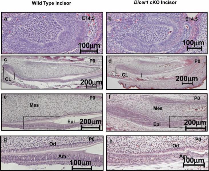
Expanded cervical loop region of incisors and repressed ameloblast differentiation in Pitx2-Cre/Dicer1 cKO. (a,b) Sagittal sections of E14.5 lower incisors, demonstrating smaller incisors in the Pitx2-Cre/Dicer1 cKO mice. (c,d) At P0, the expanded cervical loops and reduced epithelial cell differentiation were clearly observed in the mutant. Note that the cervical loop regions (CL, where tooth epithelial stem cells are located) of Pitx2-Cre/Dicer1 cKO are expanded compared with those in WT mice (lines bracket the CL). (e,g) In the WT lower incisor, tall and polarized ameloblast cells are present on the labial side. (f,h) In the Dicer1 cKO lower incisor, ameloblast cells are short and poorly polarized. (e,f) Higher magnification of (c,d). (g,h) Higher magnification of (e,f) boxed region. CL, cervical loop; Mes, mesenchyme; Epi, epithelium; Od, odontoblasts; Am, ameloblasts.

