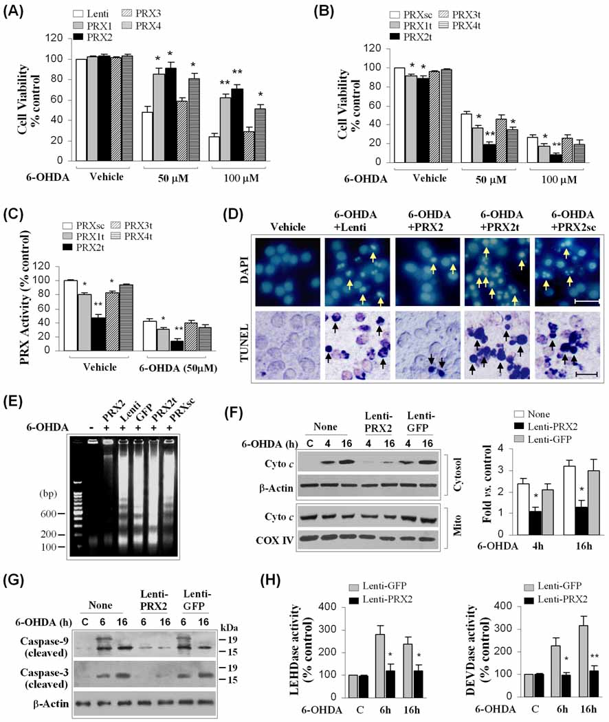
Fig. 1. Cellular 2-Cys PRXs protect against 6-OHDA neurotoxicity in MN9D cells.
(A) Neuronal differentiated MN9D cells were infected for 3 days with lentiviral vectors containing cDNA of human PRX1, PRX2, PRX3, PRX4 or the empty vector, and exposed to 6-OHDA at the indicated concentrations. Cell viability was determined at 24 h after 6-OHDA exposure. *p<0.05; **p<0.01 vs. empty vector controls, n=12 from 3 independent experiments. (B) MN9D cells were transfected for 3 days with lentiviral vectors containing shRNA targeting PRX1 (PRX1t), PRX2 (PRX2t), PRX3 (PRX3t), PRX4 (PRX4t), or the PRX2 scramble control sequence (PRXsc), and exposed to 6-OHDA. Cell viability was determined at 24 h after 6-OHDA exposure. *p<0.05; **p<0.01 vs. PRXsc controls, n=12 from 3 independent experiments. (C) Under the conditions of (B), total cellular PRX activity was determined at 2 h after 6-OHDA (50 µM) or vehicle. *p<0.05; **p<0.01 vs. PRXsc controls, data from 4 experiments. (D) Nuclear staining (DAPI) and TUNEL staining at 24 h after 6-OHDA (50 µM) in MN9D cells infected with lentiviral vectors for PRX2 over-expression (PRX2) or knockdown (PRX2t). Note that PRX2 over-expression attenuates, whereas PRX2 knockdown promotes, nuclear apoptosis and DNA fragmentation (arrows) following 6-OHDA exposure. (E) PRX2 over-expression attenuates, whereas PRX2 knockdown promotes, apoptotic DNA fragmentation 24 h after 6-OHDA exposure, as determined using DNA gel electrophoresis. The gel is representative of 3 experiments with similar results. (F) PRX2 over-expression attenuates cytochrome c release at 4 and 16 h after 6-OHDA (50 µM) exposure, determined by subcellular fractionation and immunoblotting. The graph in the right panel illustrates the cytosolic cytochrome c after 6-OHDA in cells infected with Lenti-PRX2 or the control vector Lenti-GFP. *p<0.05 vs. non-infected controls, n=4/condition. (G–H) PRX2 over-expression attenuates the activation of caspase-9 and caspase-3 at 6 and 16 h after 6-OHDA (50 µM) exposure, determined by immunoblotting against cleaved caspases (G) and peptide substrate-based protease activity assays (LEHD for caspase-9, DEVD for caspase-3), respectively (H). *p<0.05; **p<0.01 vs. Lenti-GFP-infected controls, n=4/condition.

