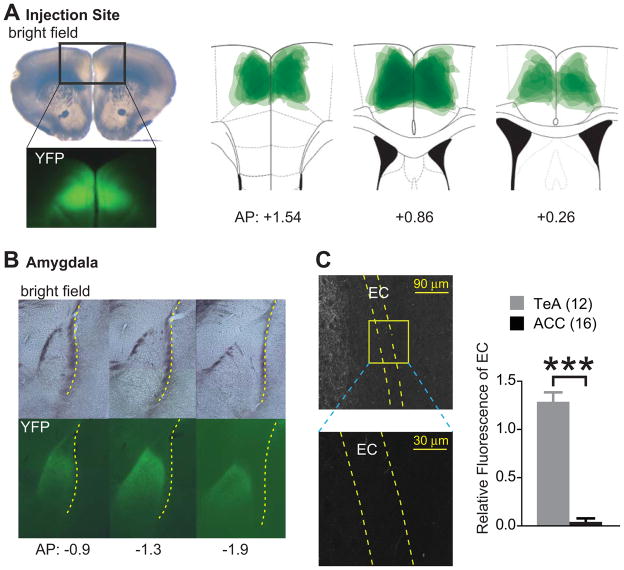
Figure 4.
ChR2-Venus expressing axons from ACC innervate LA, but bypass EC. (A) A representative slice containing ACC infected with ChR2-AAV. Upper left: bright field image, lower leftt: fluorescence image (YFP) of the corresponding rectangle area. Right: overlay of YFP expressing areas at injection sites from 8 animals. (B) Coronal sections of LA under bright field illumination (top) and fluorescence (YFP) (bottom). Numbers at bottom indicate distance from the bregma. Yellow dotted line depicts EC. (C) Left: Confocal images of YFP expressing fibers around EC from a mouse injected with ChR2-AAV in ACC. Magnified image represents yellow rectangular areas containing LA EC. Scale bars are 90 and 30 μm. Right: Relative YFP fluorescence in EC of mice injected ChR2-AAV into either ACC or TeA. Number of animals is shown in parenthesis. ***p < 0.001. Error bars represent s.e.m.

