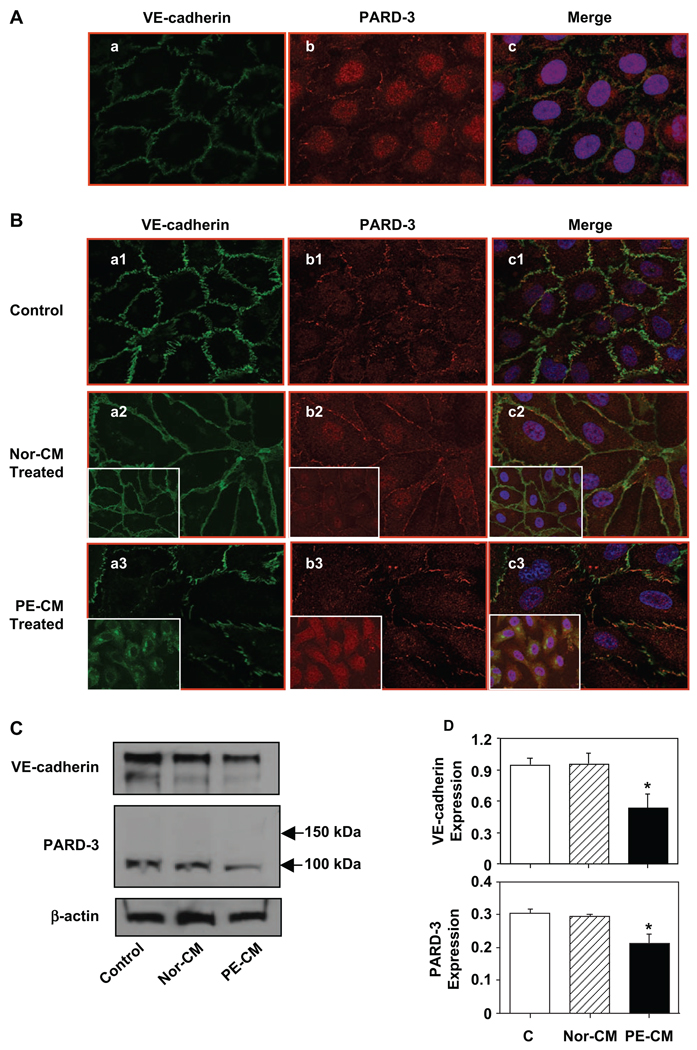
Figure 1.
Expression and distribution of VE-cadherin and partitioning defective-3 (PARD-3) in endothelial cells with or without exposure to placental conditioned medium. A, Immunofluorescent staining of VE-cadherin and PARD-3 in confluent endothelial cells (ECs); (a) VE-cadherin; (b) PARD-3; and (c) a and b merged with DAPI (nucleus) staining. Both VE-cadherin and PARD-3 locate at cell contact regions in confluent ECs. B, Immunofluorescent staining of VE-cadherin (a) and PARD-3 (b) in control cells (a1, b1, and c1) and in cells treated with normal (Nor-CM: a2, b2, and c2) and preeclamptic (PE-CM: a3, b3, and c3) placental conditioned medium (CM) for 2 hours. The inserts show cells treated with normal or PE-CM for 24 hours. No significant changes for VE-cadherin and PARD-3 expressions in cells treated with normal CM at 2 and 24 hours. Reduced and disorganized VE-cadherin and PARD-3 were seen in cells treated with PE-CM. C, Protein expressions for VE-cadherin and PARD-3 in cells treated with normal and PE placental CM by Western blot. A 100-kDa band was detected for PARD-3. Consistent with immunostaining results, both VE-cadherin and PARD-3 expressions were down-regulated in cells treated with PE-CM compared to the control cells or cells treated with normal-CM. D, Relative protein expressions for VE-cadherin and PARD-3 after normalized with actin, *P < .05. At least 3 independent assays were performed for these experiments and results are consistent.

