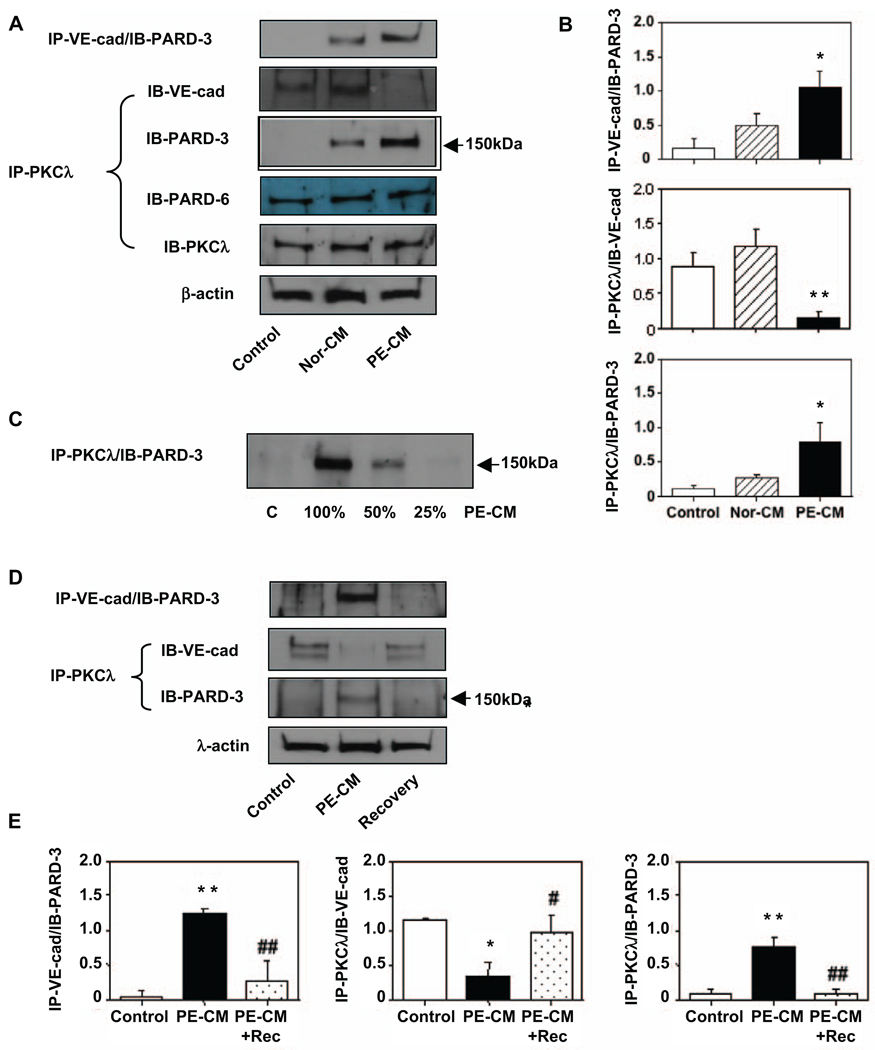
Figure 2.
Protein-protein interactions between VE-cadherin and atypical protein kinase C (aPKC)λ with partitioning defective-3 (PARD-3). A, Protein-protein interaction was evaluated in control cells and in cells that were treated with normal and preeclamptic (PE) placental conditioned medium (CM). Total cellular protein was immunoprecipitated with VE-cadherin antibody and immunoblotted with PARD-3 (IP-VE-cad/IB-PARD-3) or immunoprecipitated with aPKCλ antibody and immunobloted with VE-cad (IP-PKCλ/IB-VE-cad), PARD-3 (IP-PKCλ/ IB-PARD-3), or PARD-6 (IP-PKCλ/IB-PARD-6) and aPKCλ (IP-PKCλ/IB-PKCλ) respectively. At least 3 independent assays were performed for these experiments. These results suggest that formations of VE-cad/PARD-3 and aPKCλ/PARD-3 complexes were present in cells treated with PE-CM. In contrast, PKCλ/VE-cadherin complex was dissociated in cells treated with PE-CM. B, Bar graphs represent protein expression for IP-VE-cad/IB-PARD-3, IP-PKCλ/IB-VE-cad, and IP-PKCλ/IB-PARD-3 shown in A, *P < .05; and **P < .01 in cells treated with PE-CM vs control cells and cells treated with Nor-CM, respectively. C, Protein-protein interaction was examined in cells treated with different concentrations of PE-CM (C-0%, 100%, 50%, and 25%). Cell lysate was immunoprecipitated with aPKCλ and immunobloted with PARD-3. Formation of PKCλ/PARD-3 complex seems associated with the potency of stimuli present in the CM. D, Protein-protein interaction was determined in control cells, in cells treated with PE-CM, and after recovery. Cell lysate was immunoprecipitated with VE-cadherin or aPKCλ and immunobloted with PARD-3 or VE-cadherin, respectively. At least 3 independent assays were performed for these experiments. These data indicate that PE-CM induced VE-cad/PARD-3 and aPKCλ/PARD-3 complexes formation are no longer present after PE-CM was removed. E, Bar graphs present relatively quantitative protein expression for IP-VE-cad/IB-PARD-3, IP-PKCλ/IB-VE-cad, and IP-PKCλ/IB-PARD-3 shown in D, *P < .05 and **P < .01 for cells treated with PE-CM vs control cells; # P < .05 and ## P < .01 for cells after recovery vs cells treated with PE-CM, respectively. IP indicates immunoprecipitation; and IB = immunoblotting.

