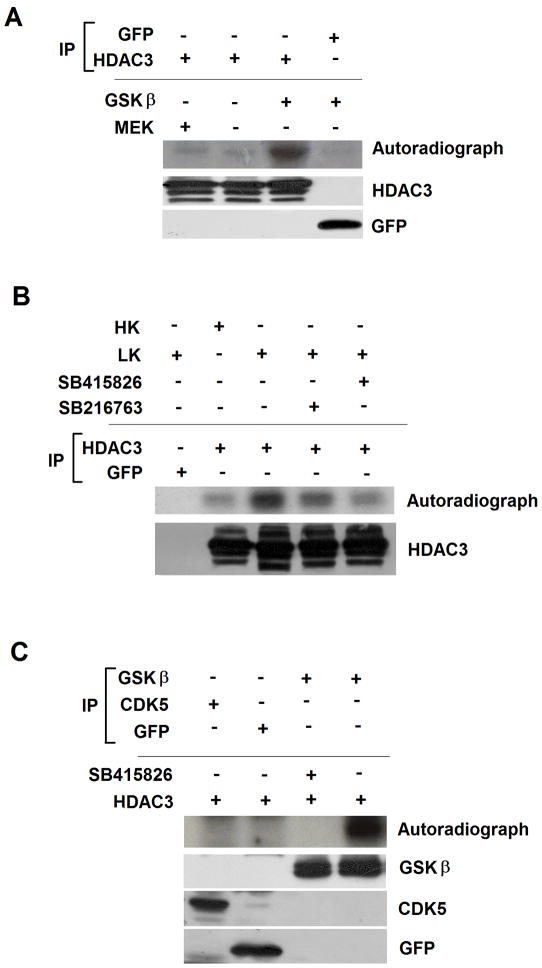
Figure 4. HDAC3 is directly phosphorylated by GSK3β.
A) Lysates from HEK293 cells transfected with either GFP or HDAC3 were immunoprecipitated using either GFP or Flag antibody. The immunpreciiated proteins were used in an in vitro kinase assay with or without active GSK3β or with active MEK. The reaction mixture was subjected to PAGE and phosphorylation evaluated by autoradiography. The lower two panels show HDAC3-Flag and GFP were immunoprecipitated in the samples used for the kinase assay.
B) CGN cultures were metabolically labeled with [32P] orthophospate in HK, LK or LK media with GSK3 inhibitors for 3 hr. Lysates from the cultures were immunoprecipitated using GFP or HDAC3 antibody and the extent of phosphorylation evaluated by PAGE and autoradiography. The lower panel shows that HDAC3 was immunoprecipitated in the samples used for the kinase assay.
C) Equal amounts of GSK3β, CDK5 or GFP immunoprecipitated from CGNs treated with LK for 6 hr were combined with HDAC3 in an in vitro kinase assays with or without a GSK3 inhibitor as indicated. HDAC3 phosphorylation was evaluated by PAGE of the reaction mixture and autoradiography. Lower panels show that similar amounts of GFP, GSK3β and CDK5 were used in the assay.

