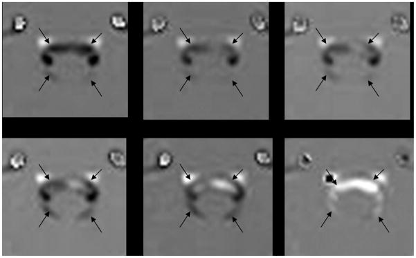
Fig 1.
From top left to lower right, PCMR images of 6 of 14 successive phases in a cardiac cycle in a patient with symptomatic Chiari I malformation and evidence of synchronous bidirectional flow. The CSF subarachnoid space is indicated with black arrows. Flow changes during these 6 phases go from a negative (black) to a positive direction (white). During the final 3 phases in the series, flow is evident in both positive and negative directions.

