
Figure 1. Characterization of Cancer Stem Cells from human HCC tissues.
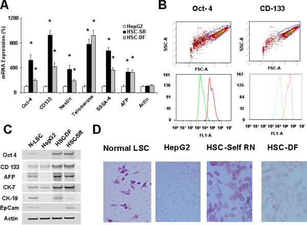
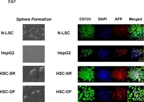
Panel A: Real-time analysis of Oct 4, CD 133, Nestin, telomerase, SSEA-4, AFP and CEA mRNA expression in HCC stem cell under self-renewal condition (HSC-SR), under differentiation (HSC-DF) and HepG2 HCC cells. All the markers are significantly up-regulated in HCC cancer stem cells. *p<0.05 when compared with HepG2 group. Panel B: FACS analysis of HCC stem cells on day 1 before cell sorting. Results are given as the percentage of Oct-4 and CD133 positive cells in the total population. In the histograms, the red line represents staining with Oct-4 or CD133 mAb, and the green lines represent the isotype control-matched mAb. Panel C: Cell lysates were obtained and immunoblotting analysis was performed for Oct-4, CD133, alpha-fetoprotein (AFP), CK-7, CK-19 and EpiCam using specific antibodies. The blots were stripped and reprobed for β-actin as a loading control and for quantitation. Panel D: StemTAG™ Alkaline Phosphatase assays performed at passage 4-6 HSCs and LSCs. HSC-SR and LSCs are maintained in an undifferentiated stage on specific culture dishes, as indicated by the high AP activity, thus confirming the self-renewal within these cells. Panel E: Formation of tumor spheres in HCC stem cells. In 1% FCS culture media 70 to 90% of the HSC-SR and HSC-DF became adherent after 7 days, with a minority of floating cells forming spheres composed of 3-5 cells. After an additional 7 days, the floating spheres were expanded to contain 15-20 HSC-SR cells, with bright appearance and sharp edge. Panel F: Immunocytochemistry for CD133 and AFP was performed in Normal liver stem cells (N-LSC), HepG2 cells, hepatocellular cancer stem cells growing under self-renewal conditions (HSC-SR) and differentiation conditions (HSC-DF). An increase in CD133 expression along with the enhanced expression of AFP is observed in HSC-SR and HSC-DF cells.