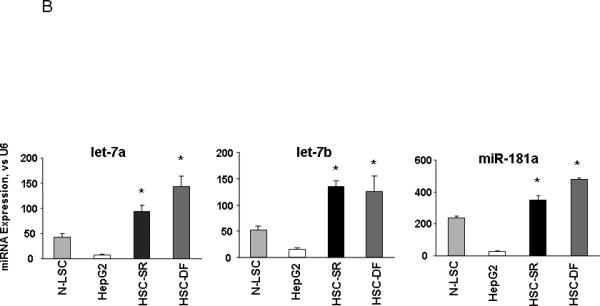
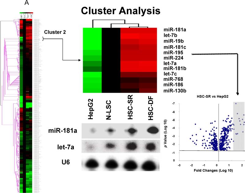
Figure 3. miRNA expression profiles in human HCC cancer stem cells (under self-renewal (SR) or differentiation (DF)), malignant and nonmalignant hepatocytes.

Panel A: miRNA was isolated and profiling by hybridization to miRNA-specific probes on epoxy-coated slides. Cluster analysis identifies a group of miRNA that are increased in expression in HCC cancer stem cells (HSC-SR and HSC-DF) and reduced in hepG2 cells when compared to normal human liver stem cells (Cluster 2). An enlarged view of this group, containing 11 miRNAs including members of let-7 and miR-181 family is shown. Lower middle panel illustrated representative Northern blot analysis of let-7a and miR-181a using total RNA isolated from four cell lines. This cluster of miRNAs are among the group, which increased in HSC-SR by 4-fold, and p<0.05 when compared to HepG2 cells (lower right panel). Panel B: The expressions of mature let-7a, let-7b and miR-181a miRNAs were validated using real-time PCR and correlated with the pattern in the miRNA array. Data represent mean ± standard error from four separate experiments.

