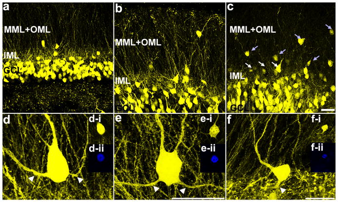
Figure 3.
Granule cell dispersion induced by intrahippocampal injection of kainic acid. Confocal maximum projections illustrating the location of the granule cell layer (GCL), inner molecular layer (IML) and outer two-thirds of the molecular layer (MML+OML) in a (a) control mouse, (b) 1wk-IHpKA mouse and (c) 2wk-IHpKA mouse. Note the greater numbers of ectopic granule cells in the inner (white arrows) and outer two-thirds (light blue arrows) of the dentate molecular layers in the ipsilateral hemisphere of a SE-2wk mouse. Scale bar = 30 μm. d-f: Examples of YFP-expressing dentate granule cells with recurrent basal dendrites (white arrowheads) from the ipsilateral hemisphere of KA-SE mice. Granule cells are located in the molecular layer of the dentate gyrus. The insets in (d-f) are optical sections through the granule cell soma showing the colocalization of YFP (d-i, e-i, f-i) and Prox1 (d-ii, e-ii, f-ii). Scale bar in e applies to d as well and = 20 μm. Scale bar for f = 20 μm.

