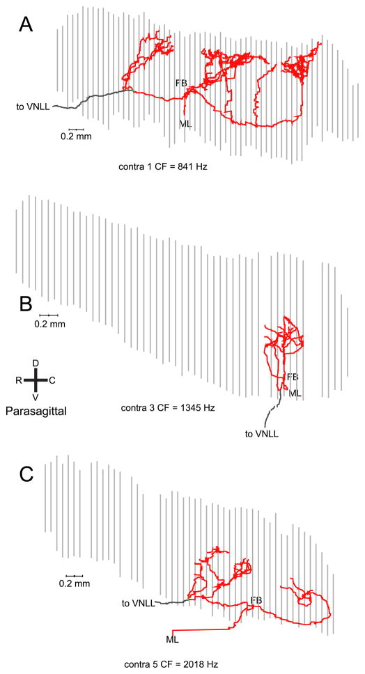
Fig. 4.
Parasagittal view of 3 more contralateral projections with a less clear cut delay line configuration. In all cases, the first branchpoint (FB) is located near the center of the rostrocaudal range of EPs. The orientation shown in panel B applies to all panels. The color convention is as in Fig. 3.

